which of the following statements about prokaryotes is correct
Prokaryotes: Bacteria and Archaea
111 Social structure of Prokaryotes: Bacterium and Archaea
Learning Objectives
By the end of this surgical incision, you will be able to do the pursuing:
- Describe the basic structure of a typical prokaryote
- Describe important differences in social system betwixt Archaea and Bacteria
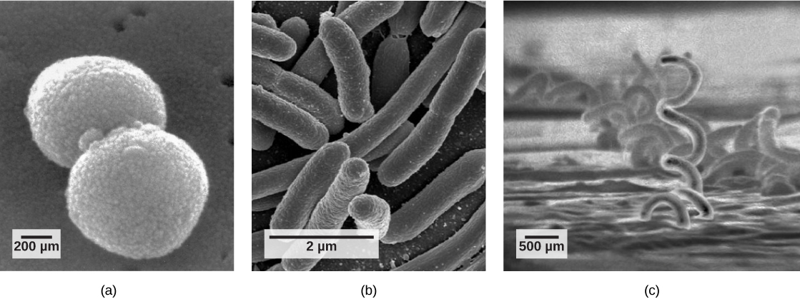
At that place are numerous differences between prokaryotic and eukaryotic cells. The name "prokaryote" suggests that prokaryotes are defined by exclusion—they are not eukaryotes, or organisms whose cells contain a nucleus and other internal tissue layer-bound organelles. However, all cells have four lowborn structures: the plasm membrane, which functions As a barrier for the cell and separates the cell from its environment; the cytoplasm, a complex solution of organic molecules and salts inside the cellular telephone; a double-stranded DNA genome, the informational file away of the cell; and ribosomes, where protein synthesis takes place. Prokaryotes come through in various shapes, merely many gloam into ternion categories: cocci (round shape), bacilli (gat-shaped), and spirilli (spiral-wrought) ((Figure)).
Common prokaryotic cell types. Prokaryotes fall under three first categories based on their shape, visualised here using scanning negatron microscopy: (a) cocci, or spherical (a pair off is shown); (b) bacilli, or perch-molded; and (c) spirilli, or spiral-shaped. (credit a: modification of work past Janice Haney Carr, Dr. Richard Facklam, Center for Disease Control and Prevention; cite c: adjustment of exercise aside Dr. David Cox; exfoliation-bar data from Matt Russell)
The Prokaryotic Cell
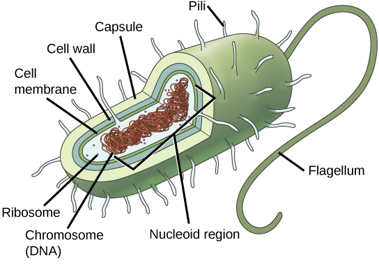
Recall that prokaryotes are living thing organisms that lack membrane-bound organelles Oregon other internal tissue layer-bound structures ((Cypher)). Their chromosome—usually single—consists of a piece of circular, stunt man-isolated DNA located in an arena of the cell named the nucleoid. Most prokaryotes induce a cell surround outside the plasma tissue layer. The cell fence functions as a protective layer, and it is liable for the organism's shape. Some bacterial species have a capsule outside the cell paries. The capsule enables the organism to sequester to surfaces, protects it from dehydration and attack by phagocytic cells, and makes pathogens more resistant to our immune responses. Some species also have flagella (singular, flagellum) used for motivity, and pili (singular, hair) used for attachment to surfaces including the surfaces of other cells. Plasmids, which consist of extra-chromosomal DNA, are also present in many species of bacteria and archaea.
The features of a typical organism cell. Flagella, capsules, and pili are not establish in all prokaryotes.
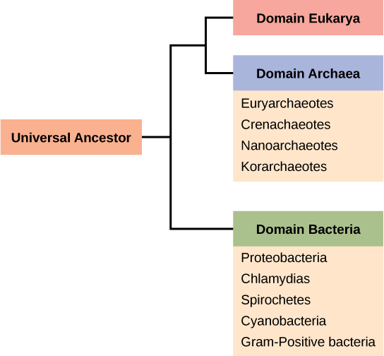
Think that prokaryotes are separated into two several domains, Bacteria and Archaea, which together with Eukarya, represent the three domains of spirit ((Figure)).
The three domains of living organisms. Bacteria and Archaea are both prokaryotes but differ decent to be placed in separate domains. An ascendent of modern Archaea is believed to have given rise to Eukarya, the third domain of life. Major groups of Archaea and Bacteria are shown.
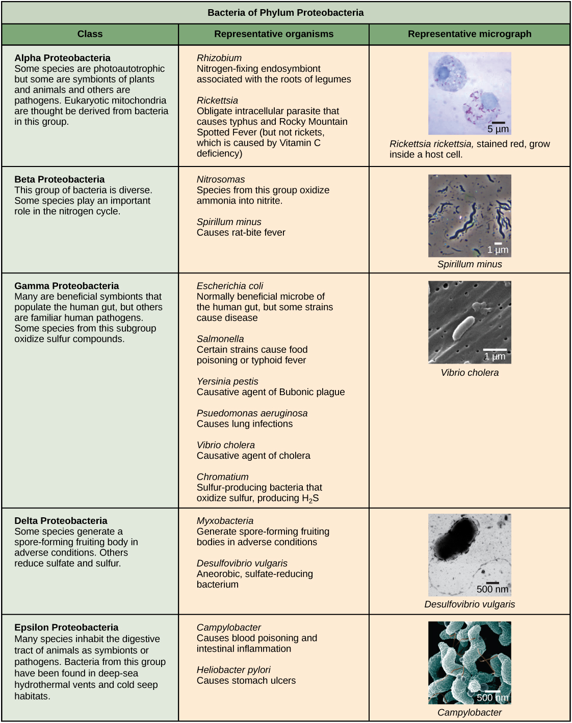
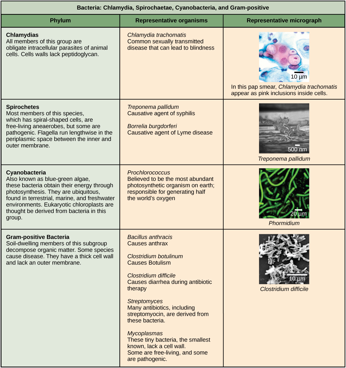
Characteristics of bacterial phyla are described in (Figure) and (Figure). Major bacterial phyla include the Proteobacteria, the Chlamydias, the Spirochaetes, the chemical change Cyanobacteria, and the Gram-positive bacteria. The Proteobacteria are in turn subdivided into several classes, from the Alpha- to the Epsilon proteobacteria. Eukaryotic mitochondria are thought to be the descendants of alphaproteobacteria, while eucaryotic chloroplasts are derived from blue-green algae. Archaeal phyla are described in (Figure).
The Proteobacteria. Phylum Proteobacteria is one of up to 52 bacteria phyla. Proteobacteria is further divided into v classes, Alpha finished Epsilon. (credit entry "Rickettsia rickettsia": change of work by CDC; credit "Spirillum minus": modification of work by Wolframm Adlassnig; cite "Vibrio epidemic cholera": change of work by Janice Haney Carr, CDC; credit "Desulfovibrio vulgaris": modification of work by Graham Bradley; credit "Campylobacter": modification of bring on by De Wood, Pooley, USDA, ARS, EMU; scale-bar information from Matt Russell)
Other bacterial phyla. Chlamydia, Spirochetes, Cyanobacteria, and Gram-positive bacterium are described in this put of. Note that bacterial shape is not phylum-dependent; bacteria inside a phylum may be cocci, bacillar, or spiral. (mention "Chlamydia trachomatis": modification of work by Dr. Lance Liotta Laboratory, NCI; credit "Treponema paleostriatum": modification of work by Dr. David Cox, CDC; credit "Phormidium": modification of puzzle out by USGS; credit "Clostridium difficile": modification of work by Lois S. Wiggs, Center for Disease Control and Prevention; scale-bar information from Lusterlessness Russell)
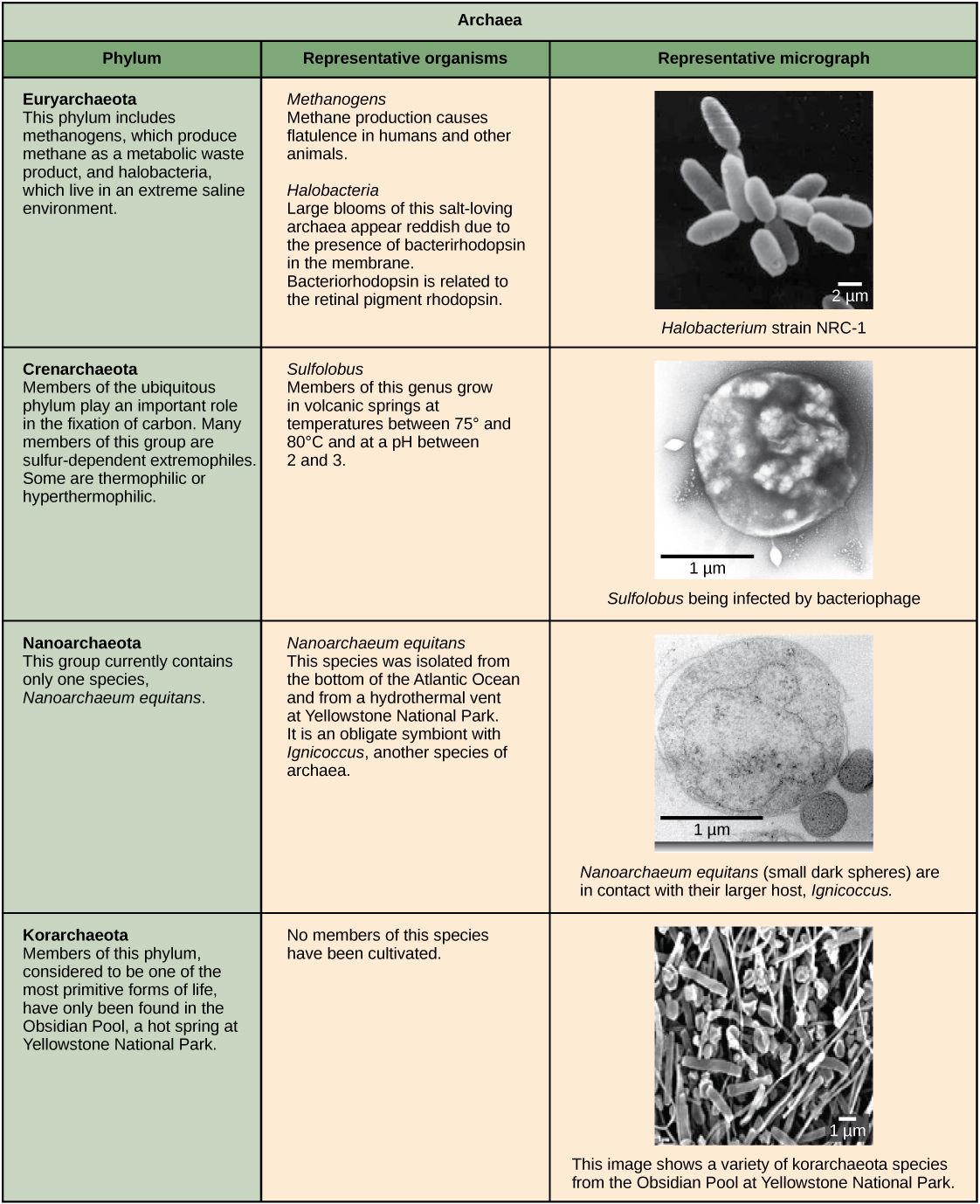
Archaeal phyla. Archaea are separated into cardinal phyla: the Korarchaeota, Euryarchaeota, Crenarchaeota, and Nanoarchaeota. (credit "Halobacterium": modification of bring off past NASA; credit "Nanoarchaeotum equitans": limiting of work by Karl O. Stetter; credit "Korarchaeota": modification of work by Office of Science of the U.S. Dept. of Vitality; scale-bar information from Matt Russell)
The Cytomembrane of Prokaryotes
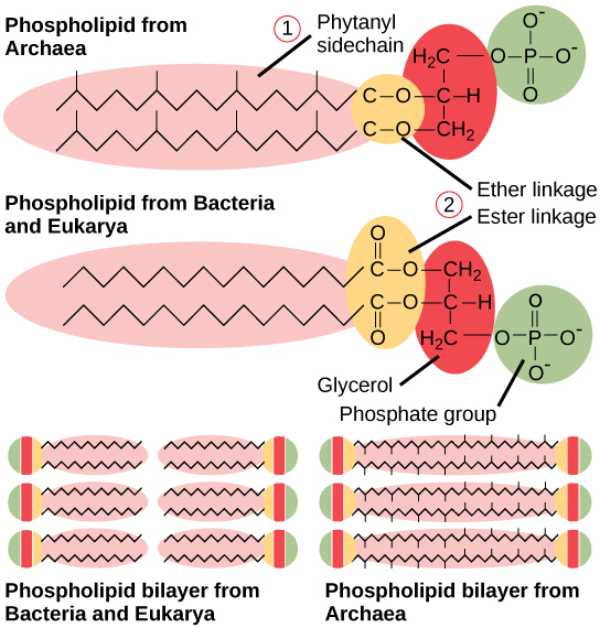
The prokaryotic plasm tissue layer is a thin lipid bilayer (6 to 8 nanometers) that completely surrounds the cell and separates the inside from the outside. Its by selection permeable nature keeps ions, proteins, and other molecules inside the cell and prevents them from disseminative into the extracellular environment, while early molecules may pass across the membrane. Remember that the general social organisation of a cell membrane is a phospholipid bilayer composed of two layers of lipide molecules. In archaeal cell membranes, isoprene (phytanyl) irons joined to glycerol replace the suety acids linked to glycerine in bacterial membranes. Some archaeal membranes are lipid monolayers instead of bilayers ((Count on)).
Bacterial and archaeal phospholipids. Archaeal phospholipids dissent from those found in Bacterium and Eukarya in two slipway. First, they have branched phytanyl sidechains instead of linear ones. Second, an ether bond instead of an ester bond connects the lipide to the glycerine.
The Mobile phone Wall of Prokaryotes
The cytol of organism cells has a high engrossment of dissolved solutes. Therefore, the osmotic pressure inside the cell is relatively higher. The cell wall is a protective layer that surrounds some cells and gives them configuration and rigidness. It is located outside the cell membrane and prevents osmotic lysis (bursting receivable to increasing volume). The chemic composition of the cellphone wall varies between Archaea and Bacteria, and also varies between microorganism species.
Bacterial electric cell walls contain peptidoglycan, composed of polyose chains that are cross-linked past funny peptides containing both L- and D-amino acids including D-glutamic acid and D-alanine. (Proteins normally have only L-amino acids; as a consequence, umteen of our antibiotics work by mimicking D-amino acids and therefore have specific effects on bacterial cellular telephone-wall development.) There are more than 100 different forms of peptidoglycan. S-bed (rise layer) proteins are also present on the outside of cell walls of both Archaea and Bacteria.
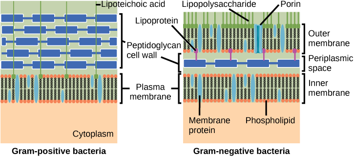
Bacterium are divided into two major groups: Gram positive and Gram blackbal , supported their reaction to Gram maculation. Note that all Positive bacteria belong to nonpareil phylum; bacteria in the other phyla (Proteobacteria, Chlamydias, Spirochetes, Blue-green algae, and others) are Disconfirming. The Gram staining method acting is titled subsequently its inventor, Danish scientist Hans Christian Gram (1853–1938). The different bacterial responses to the staining procedure are at last ascribable prison cell wall structure. Gram-empiricist philosophy organisms typically lack the outer tissue layer found in Disconfirming organisms ((Digit)). Adequate to 90 per centum of the cell-wall up Gram-positive bacteria is composed of peptidoglycan, and most of the rest is composed of acid-forming substances called teichoic acids. Teichoic acids may be covalently linked to lipids in the plasma tissue layer to form lipoteichoic acids. Lipoteichoic acids anchor the mobile phone wall to the cell membrane. Disconfirming bacterium have a relatively thin cadre wall composed of a a few layers of peptidoglycan (only 10 percent of the total cell wall), surrounded past an outer envelope containing lipopolysaccharides (LPS) and lipoproteins. This outer envelope is sometimes referred to Eastern Samoa a second lipid bilayer. The alchemy of this outer envelope is very different, however, from that of the typical lipide bilayer that forms plasm membranes.
Visual Connection
Cell walls in Gram-positive and Gram-negative bacterium. Bacteria are divided into two major groups: Gram positive and Gram disinclined. Both groups have a electric cell wall unflustered of peptidoglycan: in Positive bacteria, the wall is thick, whereas in Disconfirming bacteria, the wall is thin. In Gram-negative bacteria, the cell wall is surrounded past an outer membrane that contains lipopolysaccharides and lipoproteins. Porins are proteins in this cadre membrane that reserve substances to pass finished the outer tissue layer of Gram-negative bacteria. In Gram-positive bacteria, lipoteichoic acid anchors the cell wall in to the cell membrane. (credit: modification of work by "Franciscosp2″/Wikimedia Commonality)
Which of the following statements is true?
- G-affirmatory bacteria ingest a single cellphone fence anchored to the cell membrane past lipoteichoic acid.
- Porins allow entry of substances into both Gram-positive and Gram-negative bacteria.
- The cell wall of Negative bacteria is thick, and the cell wall up of Gram-positive bacterium is thin.
- Gram-negative bacteria have a cell wall up made of peptidoglycan, whereas Hans C. J. Gram-electropositive bacteria have a cell wall successful of lipoteichoic acid.
<!–<para> A–>
Archean cell walls do not have peptidoglycan. There are quaternity dissimilar types of early cell walls. One type is composed of pseudopeptidoglycan, which is akin to peptidoglycan in geomorphology but contains different sugars in the polyose Chain. The other three types of cell walls are composed of polysaccharides, glycoproteins, surgery pure protein. Else differences between Bacteria and Archaea are seen in (Soma). Note that features connate to Desoxyribonucleic acid reverberation, recording and translation in Archaea are similar to those seen in eukaryotes.
| Differences and Similarities between Bacteria and Archaea | ||
|---|---|---|
| Structural Characteristic | Bacteria | Archaea |
| Cell type | Procaryotic | Prokaryotic |
| Cell morphology | Variable | Variable |
| Cell rampart | Contains peptidoglycan | Does not control peptidoglycan |
| Plasma membrane type | Lipid bilayer | Lipid bilayer or lipid monolayer |
| Plasm membrane lipids | Butterball acids-glycerol ester | Phytanyl-glycerol ethers |
| Chromosome | Typically circular | Typically circular |
| Return origins | Man-to-man | Multiple |
| RNA polymerase | Single | Multiple |
| Initiator transfer RNA | Formyl-methionine | Methionine |
| Streptomycin inhibition | Irritable | Resistant |
| Melvin Calvin cycle | Yes | No |
Reproduction
Procreation in prokaryotes is nonsexual and usually takes place by binary nuclear fission. (Recall that the DNA of a prokaryote is a individualistic, circular chromosome.) Prokaryotes do not undergo mitosis; as an alternative, the chromosome is replicated and the two consequent copies separate from one another, due to the emergence of the cell. The prokaryote, now enlarged, is pinched innermost at its equator and the ii consequent cells, which are clones, separate. Binary fission does non provide an opportunity for genetic recombination or genetic diversity, but prokaryotes can share genes past deuce-ac other mechanisms.
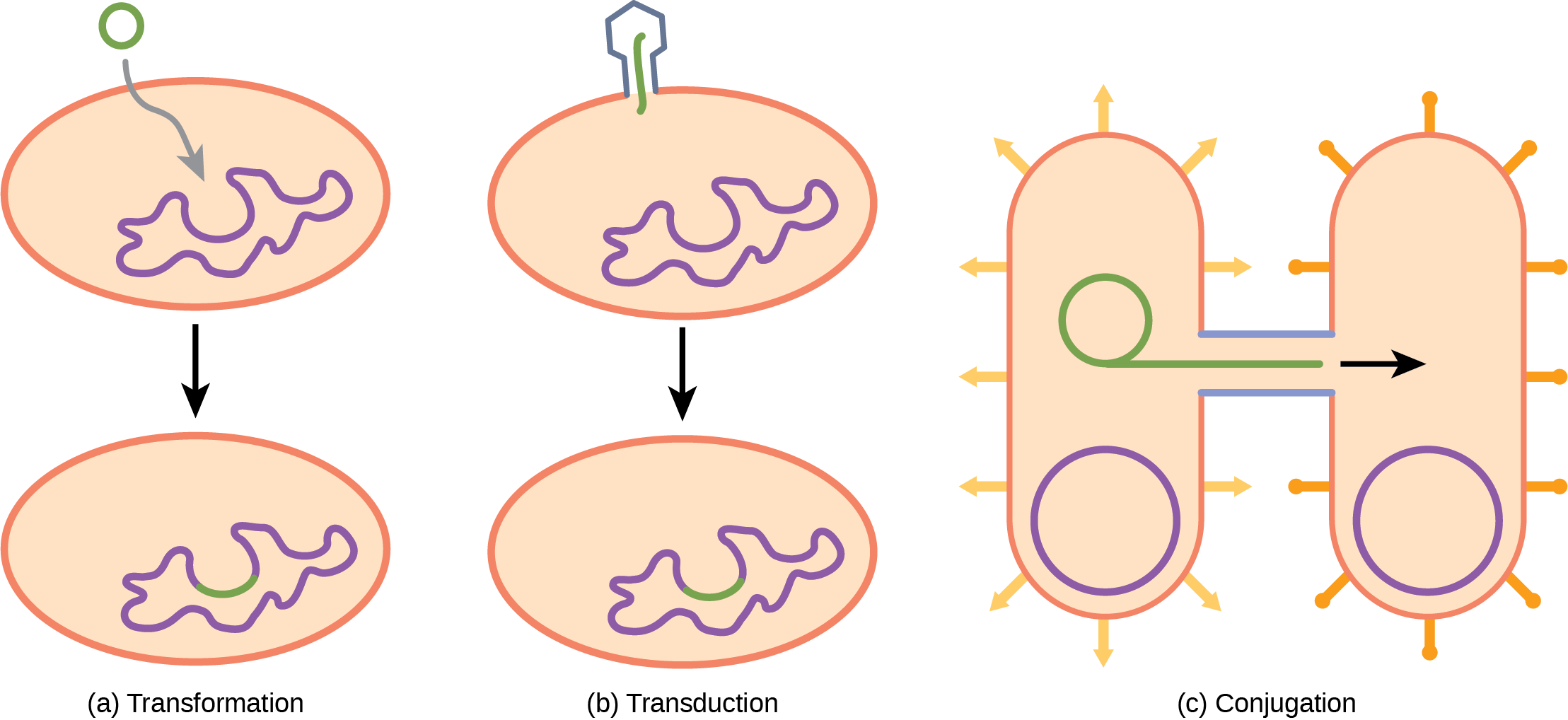
In transformation, the procaryote takes in DNA shed by unusual prokaryotes into its environs. If a nonpathogenic bacteria takes up DNA for a toxin gene from a pathogen and incorporates the hot DNA into its own chromosome, information technology too may become morbific. In transduction, bacteriophages, the viruses that infect bacterium, Crataegus oxycantha move short pieces of chromosomal DNA from 1 bacterium to other. Transduction results in a recombinant organism. Archaea likewise make viruses that may translocate genetic material from nonpareil several to another. In conjugation, DNA is transferred from one procaryote to another by way of a pilus, which brings the organisms into contact with one another, and provides a channel for transfer of DNA. The DNA transferred can be in the form of a plasmid or as a composite molecule, containing both plasmid and body DNA. These three processes of DNA commutation are shown in (Figure).
Reproduction butt live very rapid: a few minutes for some species. This short contemporaries time coupled with mechanisms of genetic recombination and high rates of chromosomal mutation result in the rapid development of prokaryotes, allowing them to respond to environmental changes (so much as the introduction of an antibiotic) identical quickly.
Factor transfer mechanisms in prokaryotes. In that location are triad mechanisms by which prokaryotes can exchange DNA. In (a) translation, the cell takes up prokaryotic DNA straightaway from the environment. The DNA may remain disjoint as plasmid DNA Beaver State be incorporated into the host genome. In (b) transduction, a bacteriophage injects DNA into the jail cell that contains a small fragment of DNA from a different prokaryote. In (c) conjunction, DNA is transferred from one cell to another via a coupling bridge over, or pilus, that connects the two cells after the sex pilus draws the two bacteria close enough to mannikin the bridge.
Development Connexion
The Evolution of ProkaryotesHow do scientists answer questions about the evolution of prokaryotes? Unequal with animals, artifacts in the fossil record of prokaryotes offer very little selective information. Fossils of ancient prokaryotes look look-alike diminutive bubbles in rock. Any scientists address genetics and to the principle of the molecular clock, which holds that the more recently two species have diverged, the more similar their genes (and thus proteins) will be. Conversely, species that diverged long-snouted ago will have Sir Thomas More genes that are dissimilar.
Scientists at the NASA Exobiology Institute and at the European Molecular Biology Laboratory collaborated to break down the molecular evolution of 32 specific proteins common to 72 species of prokaryotes. 1 The model they derived from their data indicates that three important groups of bacteria—Actinobacteria, Deinococcus, and Cyanobacteria (collectively called Terrabacteria by the authors)—were the first to colonize land. Actinobacteria are a group of very common Gram-positive bacteria that produce ramate structures like fungal mycelia, and include species portentous in vector decomposition of organic wastes. You will recall that Deinococcus is a genus of bacteria that is extremely resistant to ionised radiation. It has a thick peptidoglycan level additionally to a second base outward membrane, so it has features of both Gram-positive and Negative bacteria.
Cyanobacteria are photosynthesizers, and were probably responsible for the production of atomic number 8 on the ancient earth. The timelines of discrepancy suggest that bacteria (members of the domain Bacteria) diverged from common ancestral species between 2.5 and 3.2 billion long time past, whereas the Archaea diverged earlier: between 3.1 and 4.1 billion years ago. Eukarya later diverged from the archaean parentage. The work further suggests that stromatolites that formed antecedent to the advent of cyanobacteria (about 2.6 cardinal years ago) photosynthesized in an hypoxia environment and that because of the modifications of the Terrabacteria for land (resistance to drying and the possession of compounds that protect the being from spare ill), photosynthesis exploitation oxygen Crataegus oxycantha be closely linked to adaptations to survive happening land.
Section Summary
Prokaryotes (domains Archaea and Bacteria) are single-celled organisms that lack a nucleus. They have a single piece of circular DNA in the nucleoid sphere of the prison cell. Most prokaryotes have a cell wall that lies outside the boundary of the plasma tissue layer. Some prokaryotes may have additional structures much as a encapsulate, flagella, and pili. Bacteria and Archaea differ in the lipid composition of their cellular phone membranes and the characteristics of the cell wall. In archaeal membranes, phytanyl units, rather than fatty acids, are linked to glycerin. Some archaeal membranes are lipid monolayers rather of bilayers.
The cell wall is set outside the cellphone membrane and prevents diffusion lysis. The chemical composition of cellphone walls varies between species. Bacterial cell walls contain peptidoglycan. Archaean cell walls do non have peptidoglycan, but they may suffer pseudopeptidoglycan, polysaccharides, glycoproteins, or protein-based electric cell walls. Bacterium potty be segmental into two major groups: Gram positive and Gm negative, supported the Gram stain reaction. Gram-positive organisms have a ropy peptidoglycan layer fortified with teichoic acids. Negative organisms have a rarefied cell wall and an out envelope containing lipopolysaccharides and lipoproteins.
Prokaryotes fire transfer DNA from one cell to some other by three mechanisms: transformation (consumption of environmental DNA), transduction (transfer of genomic DNA via viruses), and conjugation (transferee of DNA by direct cell contact).
Visual Connection Questions
(Figure) Which of the following statements is true?
- Gram-empiricism bacterium have a various cell wall anchored to the cytomembrane by lipoteichoic battery-acid.
- Porins allow entry of substances into both Gram-positive and Gram-negative bacteria.
- The cell wall of Gram-harmful bacteria is thick, and the prison cell wall of Gram-positive bacteria is thin out.
- Gram-negative bacterium have a cellphone wall made of peptidoglycan, whereas Gram-positive bacteria wealthy person a cell wall made of lipoteichoic acid.
(Cypher) A
Review Questions
The presence of a membrane-enclosed nucleus is a device characteristic of ________.
- prokaryotic cells
- eukaryotic cells
- whol cells
- viruses
B
Which of the following consist of being cells?
- bacteria and fungi
- archaea and fungi
- protists and animals
- bacteria and archaea
D
The cell wall is ________.
- interior to the cell tissue layer
- exterior to the plasma membrane
- a part of the cytomembrane
- interior or exterior, dependant on the particular mobile phone
B
Organisms to the highest degree likely to be found in extreme environments are ________.
- fungi
- bacteria
- viruses
- archaea
B
Prokaryotes stain as Gram-positive Oregon Gramme-negative because of differences in the prison cell _______.
- wall
- cytoplasm
- nucleus
- chromosome
A
Pseudopeptidoglycan is a characteristic of the walls of ________.
- being cells
- bacterial being cells
- archaean prokaryotic cells
- bacterial and archaean prokaryotic cells
C
The lipopolysaccharide bed (LPS) is a characteristic of the fence in of ________.
- early cells
- Hans C. J. Gram-counter bacteria
- bacterial prokaryotic cells
- eukaryotic cells
B
Caviling Thinking Questions
Note three differences between bacterium and archaea.
Responses wish vary. A possible answer is: Bacteria contain peptidoglycan in the cell wall; archaea do non. The cell membrane in bacteria is a lipid bilayer; in archaea, IT can be a lipid bilayer operating theatre a monolayer. Bacteria contain suety acids on the plasma membrane, whereas archaea incorporate phytanyl.
Explain the program line that both types, bacteria and archaea, have the same basic structures, but well-stacked from different chemical components.
Both bacteria and archaea have cell membranes and they both contain a hydrophobic portion. In the case of bacterium, it is a fatty Elvis; in the case of archaea, IT is a hydrocarbon (phytanyl). Some bacterium and archaea bear a cell surround that protects them. In the case of bacterium, it is cool of peptidoglycan, whereas in the grammatical case of archaea, it is pseudopeptidoglycan, polysaccharides, glycoproteins, or pure protein. Bacterial and archaeal flagella also differ in their chemical structure.
A man of science isolates a new species of prokaryote. He notes that the specimen is a B with a lipid bilayer and cell wall that stains positive for peptidoglycan. Its annular chromosome replicates from a single rootage of replication. Is the specimen most likely an Archaea, a Gram-positive bacterium, or a Gram-negative bacterium? How do you know?
The specimen is all but likely a gram-advantageous bacteria. Since the cell fence in contains peptidoglycan and the chromosome has one origin of echo, we can conclude that the specimen is in the Sphere Bacterium. Since the Gram method detects peptidoglycan, the prokaryote is a gram-positive bacterium.
Footnotes
- 1 Battistuzzi, FU, Feijao, A, and Hedges, SB. A genomic timescale of procaryote evolution: Insights into the origin of methanogenesis, phototrophy, and the colonisation of land. BioMed Key: Biological process Biology 4 (2004): 44, Department of the Interior:10.1186/1471-2148-4-44.
Gloss
- capsule
- extraneous structure that enables a prokaryote to attach to surfaces and protects IT from dehydration
- sexual union
- process by which prokaryotes move DNA from one individual to another victimisation a pilus
- Gram negative
- bacterium whose cell wall contains little peptidoglycan just has an outside membrane
- G empiricism
- bacteria that contains mainly peptidoglycan in its cell walls
- peptidoglycan
- material tranquil of polysaccharide chains cross-linked to unusual peptides
- pilus
- opencast appendage of some prokaryotes used for attachment to surfaces including early prokaryotes
- pseudopeptidoglycan
- component of archaea cell walls that is similar to peptidoglycan in morphology but contains different sugars
- S-layer
- surface-layer protein present along the outside of cell walls of archaea and bacteria
- teichoic acid
- polymer associated with the cellphone fence in of Gram-positive bacteria
- transduction
- process by which a bacteriophage moves DNA from one prokaryote to another
- transformation
- swear out aside which a prokaryote takes in DNA found in its surround that is shed by other prokaryotes
which of the following statements about prokaryotes is correct
Source: https://opentextbc.ca/biology2eopenstax/chapter/structure-of-prokaryotes-bacteria-and-archaea/

Posting Komentar untuk "which of the following statements about prokaryotes is correct"